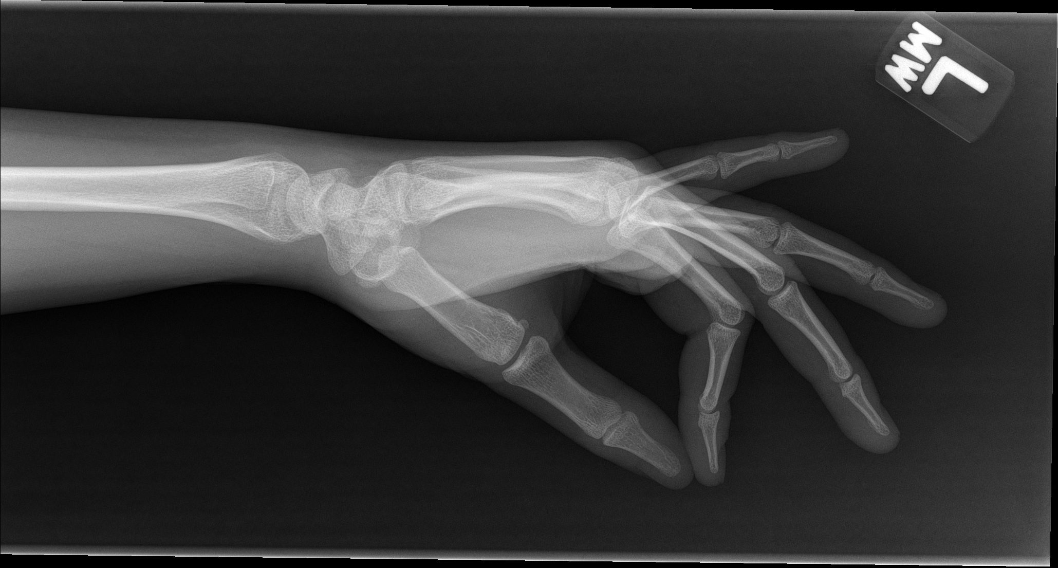
X-Ray Scans of My Ganglion Cyst
Before I was diagnosed with JRA, I had a range of other problems with the joints/bones in my body. From dancing ballet since I was 4 years old, I have two bunions— the normal one on my big toe and a tailor’s bunion on my small toe— on both feet. I also have an extra bone in the instep of my right foot, known as an extra navicular bone and pictured below:
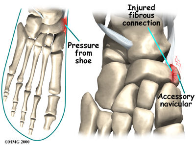
After I twisted my ankle in elementary school, I damaged the ligaments holding my extra bone in place and the pesky little bone began to rub painfully against my posterior tibial tendon. This condition is known as accessory navicular syndrome (ANS). It was hard for me to deal with ANS as an athlete because I was always moving and placing stress on my foot. Specifically for dance and figure skating, landing jumps requires our feet to absorb an impact of up to 14 times our body weight! The one podiatrist (fancy word for foot doctor) I saw suggested surgery to remove the bone, but that meant I would be unable to walk normally for more than a year. I ended up wearing arch support bands and orthopedic shoe inserts in all my shoes (including my figure skates). With the added help of time and physical therapy, my ligaments have slowly healed, and I’ve now outgrown ANS. The extra bone is still in my foot, but apparently ANS is only ANS if it hurts.
More recently, after my arthritis diagnosis I’ve noticed a lump at the base of my thumb, in the area where my palm meets my wrist. Prior to this, my arthritis did not affect my wrists, so I thought the swollen area was just a natural progression of my condition. However, it hurt more than normal, so I had x-rays and ultrasounds done. We eventually identified the lump as a ganglion cyst. Or in my doctor’s words, “Multiloculated ganglion cyst versus ganglion of the tendon sheet at the level of the first metacarpal-carpal joint.”
It’s pictured as the small black oval haze located almost directly below my second metacarpal. Because a ganglion cyst is primarily composed of fluid, it does not show up very well on x-rays. That was part of the reason why it took multiple scans for my doctors to confirm. Ultrasounds were more useful in my case because they can better detect soft tissue. From my knowledge, a ganglion cyst is basically excess synovial joint fluid that was squished out of my joint capsule. It is non-cancerous and often resolves itself, although sometimes surgery is used to drain particularly bothersome cysts. I chose to avoid surgery, and while my cyst has shrunken in size, it is still here one year later.
It was very interesting for me to learn about the medical terminology behind the conditions that I had, and how/why they occurred. This was the first time I had x-ray pictures of my own body, so I thought I would share my experiences!
Any updates? I have also struggled with this condition. I’ve heard that it is also called a bible cyst. Have you tried slamming it with a bible to see if it will go away? What are your thoughts about this remedy?
Wow this is my first time seeing x-ray photos. Thank you for the detailed explanation of this condition
I’ve always told people that there was more wrong with me than just arthritis, and no one ever believed me. It’s great to see that you recognize that not every swollen thing in your body is automatically arthritis.